How Periodontists Use Technology To Improve Treatment Outcomes
Healthy gums protect your teeth, your comfort, and your confidence. When gum disease starts, small problems can turn into tooth loss and pain. Today, periodontists use modern tools to catch trouble early and treat it with less cutting and less guesswork. You see clearer images, shorter visits, and steadier results. Thousand Oaks dentist teams and gum specialists now rely on digital scans, 3D images, and gentle lasers to remove infection and shape damaged tissue with care. These tools help your periodontist plan each step before picking up an instrument. They also help track healing and spot small changes before they grow. You feel more in control. You get clear answers. You gain a treatment plan that fits your mouth and your daily life.
This blog explains how these tools work, why they matter, and how they can protect your smile.
Why technology matters for your gums
Gum disease often grows in silence. You may not feel pain until the damage is serious. New tools give your periodontist three key advantages.
- Find problems while they are still small.
- Treat infected tissue with less cutting.
- Watch healing and adjust care early.
These steps protect bone, teeth, and implants. They also reduce fear, time in the chair, and unplanned costs.
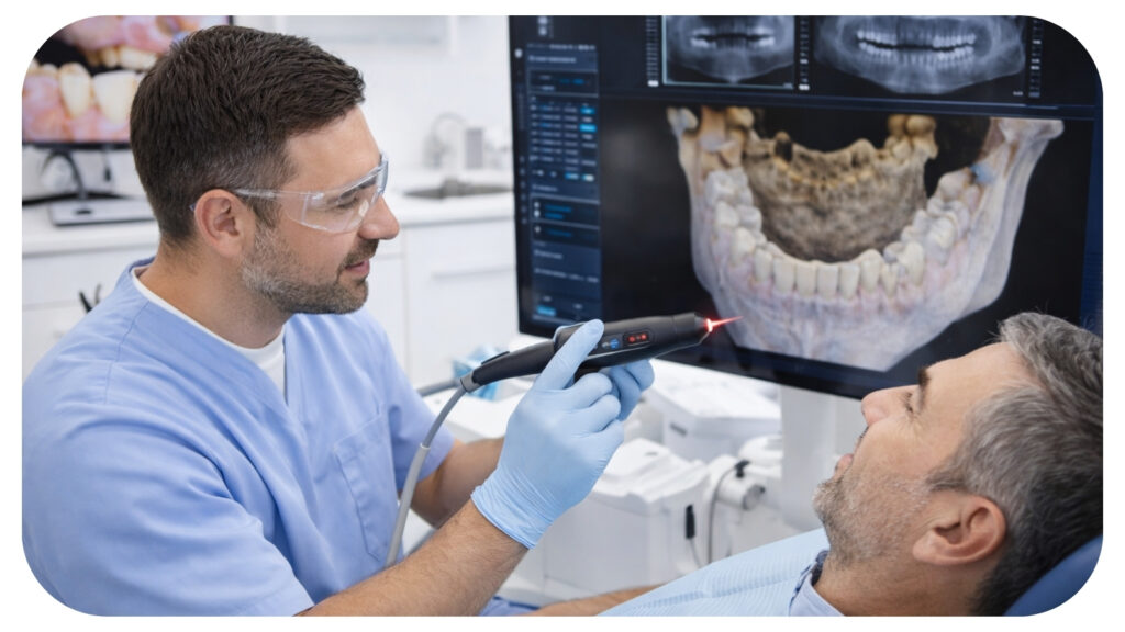
Digital X-rays and 3D scans
Digital X-rays and cone beam CT scans show bone and roots in sharp detail. They use focused beams and give clear pictures that appear on a screen in seconds.
With these images, your periodontist can:
- Measure bone loss around each tooth.
- Spot hidden infections and cysts.
- Plan safe implant positions.
- Avoid nerves and sinus spaces.
Research from the National Institutes of Health explains how 3D scans improve implant planning and lower nerve injury risk. You can read more on the NIH PubMed Central site.
Intraoral cameras and digital photos
Intraoral cameras are small cameras that fit in your mouth. They show stained plaque, gum pockets, and cracks on a screen in front of you.
This helps you:
- See the exact spots that bleed or trap food.
- Understand why a tooth or implant feels loose.
- Compare before and after views of treatment.
It also helps your periodontist document changes over time. That record supports clear decisions when gums improve or slip.
Laser treatment for gum disease
Dental lasers use focused light to remove infected tissue and clean pockets. They cut with heat and light instead of a scalpel.
With lasers your periodontist can:
- Target diseased tissue and leave healthy tissue in place.
- Reduce bleeding during and after treatment.
- Lower swelling and shorten healing time.
Many people need fewer stitches. Many also report less post-treatment soreness. This can make it easier to return to work and daily routines.
Guided surgery for implants
Guided surgery uses 3D scans and computer planning. Your periodontist plans the exact angle, depth, and position of each implant on a screen. Then a custom guide rests on your teeth or gums to direct the implant during surgery.
This method gives three clear benefits.
- More accurate placement.
- Shorter surgery time.
- Lower risk to nearby roots and nerves.
Accurate placement helps implants last longer. It also supports natural chewing and easier cleaning.
Digital impressions and custom guides
Digital impression scanners replace many trays of putty. A small wand moves over your teeth and gums and records thousands of images each second.
These scans help your periodontist and lab team to:
- Design guides for implant placement.
- Create crowns and bridges that fit on the first try.
- Avoid gagging and repeat visits for new molds.
For children and older adults, this can reduce stress and confusion. It also reduces errors from distorted putty molds.
Comparing common periodontal tools
| Tool | Main purpose | Key benefit for you | Typical use |
|---|---|---|---|
| Digital X-rays | View teeth and bone | Quick images with lower radiation than film | Check bone loss and cavities |
| 3D cone beam scan | Show full jaw in 3D | Safer implant and surgery planning | Implants and complex gum disease |
| Intraoral camera | Show close-up tooth and gum views | Clear pictures that you can see and understand | Gum exams and progress checks |
| Dental laser | Remove infected tissue | Less bleeding and shorter healing | Moderate and severe gum disease |
| Digital impression scanner | Capture tooth and gum shapes | No messy molds and fewer repeat visits | Implants, crowns, and gum graft guides |
Tracking healing with digital records
After treatment, your periodontist needs proof that your gums stay stable. Digital charts and images help track three key signs.
- Pocket depth around each tooth.
- Bone levels on X-rays.
- Bleeding and plaque scores.
Many offices use charting systems that show color-coded changes. If a pocket deepens or a site bleeds, the system flags it. Your periodontist can then adjust cleanings, home care, or medicine.
The Centers for Disease Control and Prevention explain that nearly half of adults over 30 have some form of gum disease. You can review national data on the CDC oral health page. Regular tracking helps you avoid moving into the severe group.
What this means for your daily life
Modern periodontal care is less about rescue and more about control. You gain three core protections.
- Earlier warning before damage spreads.
- Targeted treatment that protects healthy tissue.
- Ongoing checks that keep gums stable.
You still need daily brushing, flossing, and checkups. Yet technology gives your effort more strength. It turns small choices into lasting safety for your teeth, implants, and gums.

Most Inside Editorial Team
MostInside is a trusted publication helping people navigate real decisions across health, finance, career, home, relationships and technology since 2009. Straight to What Matters.


